

Understanding Tumors: Types And Impact On Muscle Function
Tumors are abnormal tissue growths classified as benign (non-cancerous) or malignant (cancerous). While benign tumors grow slowly and usually stay localized, malignant tumors can invade nearby tissues and spread throughout the body, making them more difficult to treat, especially near critical structures like muscles.
When tumors affect muscles, they can cause pain, weakness, or loss of function. Malignant tumors may compress or destroy muscle tissue, and even surgery can risk further damage if not carefully planned.
To manage tumors in muscle-rich areas, doctors must balance complete removal with preserving muscle function. MRI imaging is crucial, offering detailed views of the tumor and its interaction with nearby muscles to guide precise surgical planning and protect mobility. Resources such as those found at https://sarcomaoncology.com/ provide valuable insights into the complexities of treating tumors in these sensitive areas.
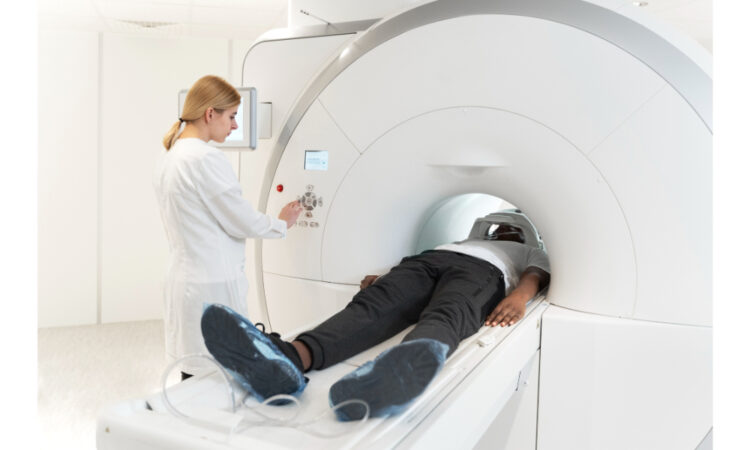
How MRI Works: A Brief Overview of the Technology
MRI uses magnetic fields and radio waves—not ionizing radiation like X-rays or CT scans—to create detailed images by targeting hydrogen atoms in water and fat. These atoms align with the magnetic field, and when disrupted by radiofrequency pulses, they emit signals that are converted into high-resolution images.
MRI excels at imaging soft tissues such as muscles, nerves, and organs, and is safer for repeated use. Different techniques enhance its capabilities: T1-weighted images show anatomy, T2-weighted images highlight inflammation, and advanced methods like DWI and fMRI provide insights into tissue properties and brain function. These features make MRI essential for tumor diagnosis and monitoring.
Advantages of MRI Over Other Imaging Techniques
MRI excels at soft tissue contrast, clearly showing muscles, nerves, and brain structures—often less visible on X-rays or CT scans—making it ideal for detecting tumors in complex areas. Its 3D imaging helps surgeons plan precise operations that remove tumors while preserving healthy tissues like muscles.
Unlike CT, MRI uses no ionizing radiation, making it safer for repeated scans, especially in long-term cancer care and for sensitive groups like pregnant women and children. This makes MRI a top choice for tumor diagnosis, planning, and monitoring.
Precision is crucial in tumor surgery, particularly near muscles and nerves, to fully remove tumors while protecting function. MRI-guided planning lets surgeons see tumor size and location, avoid critical areas, and perform tissue-sparing surgery, improving recovery and patient outcomes.
MRI in Tumor Detection and Diagnosis
Magnetic Resonance Imaging (MRI) has transformed tumor detection by providing high-resolution, non-invasive images of soft tissues. Unlike other imaging methods, MRI uses magnets and radio waves to distinguish between tissue types, making it especially effective at identifying and characterizing tumors.
Beyond detection, MRI offers detailed information about a tumor’s size, location, and its relationship to surrounding structures, particularly muscles and nearby organs. This information is especially important in surgical planning, where consulting with orthopedic surgery specialists can guide limb-sparing procedures that prioritize preserving function.
When tumors are near or within muscles, MRI’s precision helps clearly define tumor boundaries and protect healthy tissue, reducing complications and aiding recovery. In this way, MRI not only improves diagnostic accuracy but also supports better long-term outcomes.
MRI’s Contribution to Surgical Planning and Decision Making
MRI is a critical tool in tumor surgery, offering detailed, 3D images that help surgeons assess tumor size, shape, and location, along with its relationship to surrounding tissues. This clarity allows for precise planning and safer, more effective tumor removal.
Advanced techniques like functional MRI (fMRI) provide insights into areas of the brain responsible for speech, movement, and sensation, helping avoid critical regions during surgery. MRI also detects vascular involvement, aiding in blood supply management and reducing bleeding risks.
Beyond the OR, MRI supports team-based care, allowing surgeons, oncologists, and radiologists to collaborate on tailored treatment plans. By informing every step of the process, MRI strengthens both surgical precision and patient outcomes.
Future Innovations in MRI Technology for Surgical Applications
The future of MRI is marked by innovations that promise to enhance its surgical utility. Faster MRI techniques are being developed to reduce scan times without compromising image quality, improving patient comfort, minimizing motion artifacts, and increasing accessibility.
One significant advancement is intraoperative MRI with real-time imaging capabilities, which allows surgeons to assess tumor removal and surrounding structures mid-procedure. This added precision helps reduce the risk of incomplete resection while supporting safer, more effective outcomes. High-resolution imaging also plays a vital role in limb-sparing surgery by providing detailed views of tumor boundaries and adjacent healthy tissues, enabling surgeons to preserve as much function as possible.
Emerging targeted contrast agents are also transforming MRI. Designed to bind to specific cancer markers, they enable more accurate tumor detection and characterization, supporting personalized treatment.
As these technologies advance, MRI is poised to play an even more crucial role in improving surgical outcomes and shaping the future of cancer care.
Challenges and Limitations of MRI in Tumor Surgery
While MRI offers significant benefits, it has limitations. Cost and accessibility remain major barriers—many rural or underserved areas lack MRI machines, leading to unequal access to advanced cancer imaging and spine and musculoskeletal imaging. Image artifacts caused by patient movement, metal implants, or technical issues can reduce clarity and affect diagnostic accuracy. Skilled interpretation is essential to overcome these hurdles.
MRI also presents physical and safety challenges. The enclosed space can trigger claustrophobia, and strong magnetic fields make MRI unsafe for some patients with implants like pacemakers. In such cases, alternative imaging methods must be considered.
Despite these issues, MRI remains a vital tool, with its benefits far outweighing its challenges in tumor diagnosis and surgical planning.
Conclusion: The Critical Role of MRI in Enhancing Surgical Outcomes
MRI has become an indispensable tool in tumor surgery, offering unmatched clarity in visualizing soft tissues, mapping functional anatomy, and guiding precise surgical planning. Its contributions have significantly improved the detection, diagnosis, and management of tumors, enhancing surgical accuracy, preserving muscle function, and ultimately elevating patient outcomes and quality of life.
Despite limitations such as cost and accessibility, continued innovation in MRI technology, such as faster imaging, intraoperative guidance, and targeted contrast agents, promises to overcome these barriers and broaden its clinical impact. The case studies discussed illustrate MRI’s transformative role in surgical success, highlighting its essential place in contemporary medical practice.
As technology advances, MRI remains at the forefront of surgical innovation. Its ability to support precise tumor resection and comprehensive treatment planning ensures its continued relevance in delivering effective, personalized, and compassionate cancer care.